Chemotherapy is an important and often effective strategy to block or slow the growth of cancerous cells. Docetaxel is an example of a potent chemotherapeutic drug that is applied in the treatment of various malignancies, including breast, head and neck, stomach, non-small-lung, and prostate cancer.
Docetaxel is a semi-synthetic compound derived from the European yew tree that interferes with the division of chromosomes in dividing cells which results in cell death. However, not all patients treated with docetaxel show benefits; the response rates vary between 6% and 45%.
Previous research has indicated that a lack of treatment response correlates with a lack of docetaxel uptake in cancer cells. This means that delivery of the drug to the tumor is essential for it to work (treatment efficacy). Due to its significant toxicity and side effects, it is not feasible to simply increase the docetaxel dosage in patients to increase tumor exposure. Therefore, alternative tumor-delivery tools are under development to boost docetaxel levels in tumor cells, while avoiding exposure of healthy tissues.
Nanoparticles as docetaxel delivery vehicles
Developed by Cristal Therapeutics, CPC634 is an experimental nanoparticle that encapsulates docetaxel. Compared to docetaxel alone, this packaging enhances stability in the blood stream and improves tumor delivery, increasing docetaxel’s efficacy while avoiding systemic exposure. To develop a non-invasive clinical tool that can identify patients who may benefit from docetaxel delivery by CPC634, a clinical trial (NCT03712423) was initiated by Dr. Willemien Menke-van der Houven van Oordt and PhD candidate Iris Miedema. In this trial positron emission tomography/computed tomography (PET/CT) imaging was used to determine the uptake of these nanoparticles in tumor cells.
Testing PET/CT imaging feasibility
The outcome of this clinical investigation was recently published in the journal Advanced Materials. First author Iris Miedema: “It wasn’t clear if PET/CT imaging of CPC634 at a low diagnostic dose would be possible, since preclinical research indicated rapid clearance of nanoparticles from the blood by the liver. To test this, we radiolabeled CPC634 with zirconium-89. We then performed imaging with both a low dose and a higher therapeutic dose and compared the results.”
Patient stratification and therapy development
With a low dose of CPC634, the researchers were able to visualize and quantify the accumulation of the polymeric nanoparticles in solid tumors. Importantly, the low diagnostic dose accurately reflected the higher therapeutic dose imaging. “In the future, prior imaging with a low diagnostic dose may therefore help to select patients who may benefit from this treatment,” says Willemien. Patients with higher accumulation of CPC634 in the tumor are presumably more likely to benefit from the treatment than patients with low accumulation. “Moreover, it can be utilized to explore strategies to improve nanoparticle accumulation in the tumor, for example, by co-administration with vascular or other microenvironment modulators.”

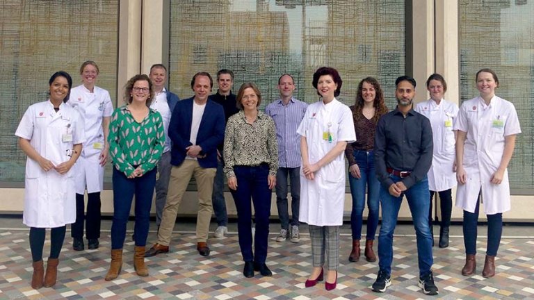
The PET imaging research team in front of the Imaging Center, Amsterdam UMC.
The promising results obtained in this clinical trial would not have been realized without team work. From left to right: Ramsha Iqbal, Hanneke Pouw, Daniëlle Vugts, Bert Windhorst, Ben Zwezerijnen, Ronald Boellaard, Willemien Menke-van der Houven van Oordt, Marc Huisman, Daniela Oprea-Lager, Jessica Wijngaarden, Maqsood Yaqub, Jelijn Knip, Iris Miedema.
For more information, read the publication.
Miedema, I. H. C., et al. (2022) PET-CT Imaging of Polymeric Nanoparticle Tumor Accumulation in Patients. Adv. Mater. 2201043. https://doi.org/10.1002/adma.202201043
Or contact Dr. Willemien Menke-van der Houven van Oordt
People at Cancer Center Amsterdam involved in the study:
Iris H.C. Miedema, Gerben J.C. Zwezerijnen, Marc C. Huisman, Ellen Doeleman, Ron H.J. Mathijssen, Twan Lammers, Qizhi Hu, Guus A. M. S. van Dongen, Cristianne J.F. Rijcken, Danielle J. Vugts, C. Willemien Menke-van der Houven van Oordt
Funding was received from Health Holland (grant) and Cristal Therapeutics.

