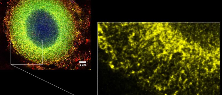
Researchers Wilma van de Berg and Tim Moors from the Amsterdam UMC joined forces with colleagues from Roche (Switzerland) and Ruhr University Bochum (Germany) to disentangle the detailed architecture of Lewy bodies in the brain with Parkinson’s disease using state-of-the-art high-resolution microscopy techniques. Their results highlight consistent structures in the composition of mature Lewy bodies, leading to new hypotheses on the formation of these features in the Parkinsonian brain.
Onion skin-like architecture of Lewy bodies
One of the major components of Lewy bodies is α-synuclein, a protein abundant in the human brain that exists in various forms. In this project – performed by dr. Tim Moors (Amsterdam UMC) and led by dr. Wilma van de Berg (Amsterdam UMC) and dr. Markus Britschgi (Roche) – different α-synuclein variants were visualized in great detail using multilabel immunofluoresecence and superresolution (STED) microscope, which allows optical imaging at a higher-than-normal resolution (e.g. ~50nm instead of 200nm). Using highly sensitive and specific antibodies developed by Roche and Prothena Biosciences Inc. and advanced microscopy techniques, the 3D architecture of Lewy bodies was further unraveled: they were found to contain different concentric ring-shaped layers, in which a core of accumulated proteins and lipids is surrounded by phosphorylated α-synuclein and structure-providing cellular components. These findings suggests that brain cells actively regulate the shape of Lewy bodies, possibly to encapsulate toxic components.
A stepwise construction of Lewy bodies
In addition to the distribution of α-synuclein variants in Lewy bodies, the authors also compared their localization outside of Lewy bodies in cells in the brains of donors with early versus late stages of Parkinson’s disease, as well as controls. The authors observed that already at early stages of Parkinson’s disease phosphorylated α-synuclein aligns in a network around the cell nucleus before Lewy bodies become apparent, suggesting that its aberrant distribution occurs before Lewy body formation. Based on their findings, the authors propose a hypothesis in which brain cells build Lewy bodies in situations where they are no longer able to recycle their debris. When lipids and proteins are accumulating, the cell might attempts to sequester them at a dedicated place – comparable to a landfill –which it eventually re-shapes and encapsulates to protect itself against harmful components. Over time the presence of this “landfill” itself might alter the normal biology of neurons.
The detailed observations in the postmortem brain of donors with Parkinson’s disease in this study provide important new insights into the consistent structured architecture of Lewy bodies and serve as an important reference for current and future model systems of Parkinson’s disease. Vice versa, testing the presented hypothesis in such experimental systems is an important future step for a better understanding of Lewy bodies in Parkinson’s disease, which is pivotal for the development of biomarkers and novel therapies that combat this devastating disease.
Read the article in Acta Neuropathologica: The subcellular arrangement of alpha-synuclein proteoforms in the Parkinson’s disease brain as revealed by multicolor STED microscopy

