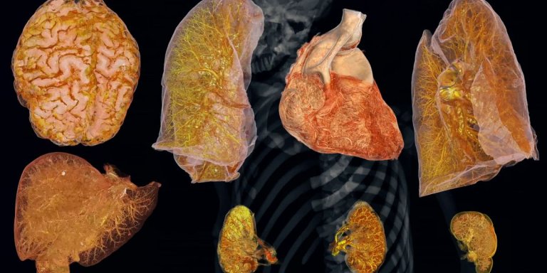
3D images have already been created of more than 50 organs, including the lungs, brain, hearts, kidneys, spleen and liver. The images are made using the organs of people who have made themselves available to science. The particle accelerator makes it possible to examine entire organs, all the way down to the cellular level. The scans have a resolution of 25 microns, which is about twenty times the resolution of a clinical CT scanner. It can then zoom in on small areas with a resolution of less than a micron, which is five hundred times the resolution of a CT scanner.
The ultimate goal is to create a global reference database of organ images, accessible to scientists and medical professionals around the world. These images are available on the HOAHub website. The images are initially intended for medical education and clinical research but will, ultimately, also benefit healthcare because the detailed images provide new insights into the causes of diseases and that in turn leads to new treatments.
The project is a collaboration of several partners and is led by University College London. Thanks to various donors, the extremely expensive European particle accelerator (about €25,000 per hour) can be used for science. Researchers can submit research proposals, if their proposal is assessed as impactful, they can access the scanner. De Bakker and seven others form the committee that assesses these applications.


