Confocal laser endomicroscopy
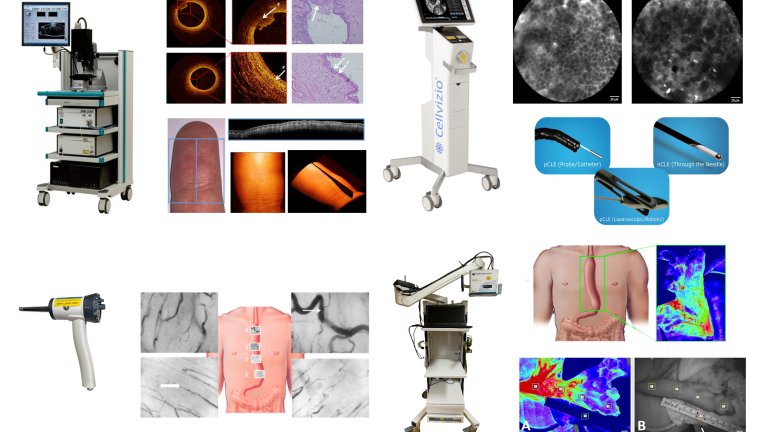
Confocal laser endomicroscopy (CLE) is an advanced imaging technique that allows real-time, high-resolution visualization of tissues at the cellular level during endoscopy or needle-based interventions. CLE is based on fluorescence and uses 488 nm light to excite either fluorescein or intrinsic auto-fluorescence (i.e. cartilage) to create in vivo confocal images. The CLE engine is interfaced with several fiber-based catheters, all using different optics depending on the required need. The imaging depth is around 60 micrometer and the field of view, depending on the used imaging catheter, ranges from 150 to 500 micrometer.
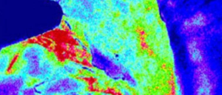
Laser speckle contrast imaging
A laser speckle contrast imager is available that allows the visualization of bulk tissue perfusion of the whole surgical field. Laser speckle contrast imaging offers rapid, full-field visualization of blood flow dynamics, enabling real-time assessment of microvascular perfusion with high spatial and temporal resolution. It is an imaging modality that visualizes the superficial blood flow based on the change in the speckle signal generated from a near infrared laser. With laser speckle contrast imaging (LSCI), a large area (10x20 cm) can be analyzed in real time giving the perfusion dynamics from tissue under investigation. The LSCI setup within the Biomedical engineering and physics department is positioned on a clinical cart, with the compact imaging head positioned conveniently on a articulating arm.
Optical coherence tomography
Different clinical and preclinical Optical Coherence Tomography (OCT) systems are available for research purposes. The OCT systems employ near infrared light, typically 800, 1060 nm or 1310 nm to create depth resolved three-dimensional microscale images of tissue based on the back scattered light. The maximal imaging depth range into tissue is usually around 2 mm with a resolution ranging from 5 to 10 micrometer. With these high resolution 3D images, quantitative and qualitative information can be extracted from the tissue. Both scattering properties as well as perfusion information can be obtained within minutes. The OCT device can be interfaced with either a handheld scanner that allows for extra corporal imaging of i.e. skin or brain perfusion or with a helical scanning in combination with a single use sterile catheter that is tailored for imaging tubular cavities such as blood vessels.
Side stream dark field
Side stream dark field (SDF) imaging is available for in-vivo visualization of microvasculature. SDF imaging offers a noninvasive window into the microcirculation, revealing superficial capillary blood flow in tissue in real-time. It is an optical technology to visualize the microcirculation of tissue. Using SDF imaging, tissue is illuminated by green light emitting diodes (LEDs). Hemoglobin in red blood cells absorbs this 530 nm light, producing a high contrast compared to surrounding tissue. To image movement of flowing RBSs, light is pulsed stroboscopically into the tissue and is detected with a camera. The SDF device is interfaced with a handheld scanner and a sterile tip that can be used in a clinical and pre-clinical environment.
Links
- Biomedical Engineering & Physics: https://bme-physics.nl/