
Physics Meets Cardiology
An MRI scanner is driven by physical principles that can be manipulated to generate tailored images. In this VIDI project, physicist PhD candidate Renske Merton programmed the scanner to capture the thoracic aorta with unprecedented anatomical detail: a spatial resolution of 1.6 millimeters in three directions, across 30 timeframes over the cardiac cycle, all within a five‑minute scan. This innovation enabled testing a key hypothesis: that aortic motion in Marfan patients is reduced compared with healthy volunteers because of increased aortic stiffness.
Beyond Aortic Diameter
Currently, Marfan patients undergo preventive surgery when the aortic root diameter exceeds 4.5–5.0 centimeters, in order to avoid type A dissection, a tearing of the ascending aortic wall. However, dissections may occur at smaller diameters, indicating that diameter alone is an imprecise marker of aortic wall degeneration. Better biomechanical markers are therefore needed to refine surgical timing in relation to the progression of aortic disease.
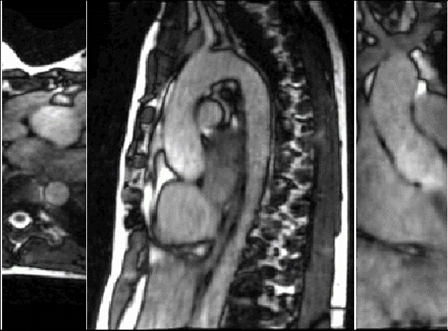 Figure 1: Detailed image 4D MRI of aortic motion
New MRI Sequence
To address this gap, Renske and MR physicist Eric Schrauben (Radiology & Nuclear Medicine) developed an MRI sequence that accurately maps aortic motion in three spatial directions over time (Figure 1). After training an nnU‑net for automatic segmentation of the aorta in each of the 30 cardiac timeframes, Renske applied image‑registration algorithms to quantify aortic displacement at every timepoint relative to diastole. With patient inclusions coordinated by PhD candidate Daan Bosshardt, the team was able to compare Marfan patients (n=82) with healthy volunteers (n=45), illustrating clear differences in aortic motion (Figure 2).
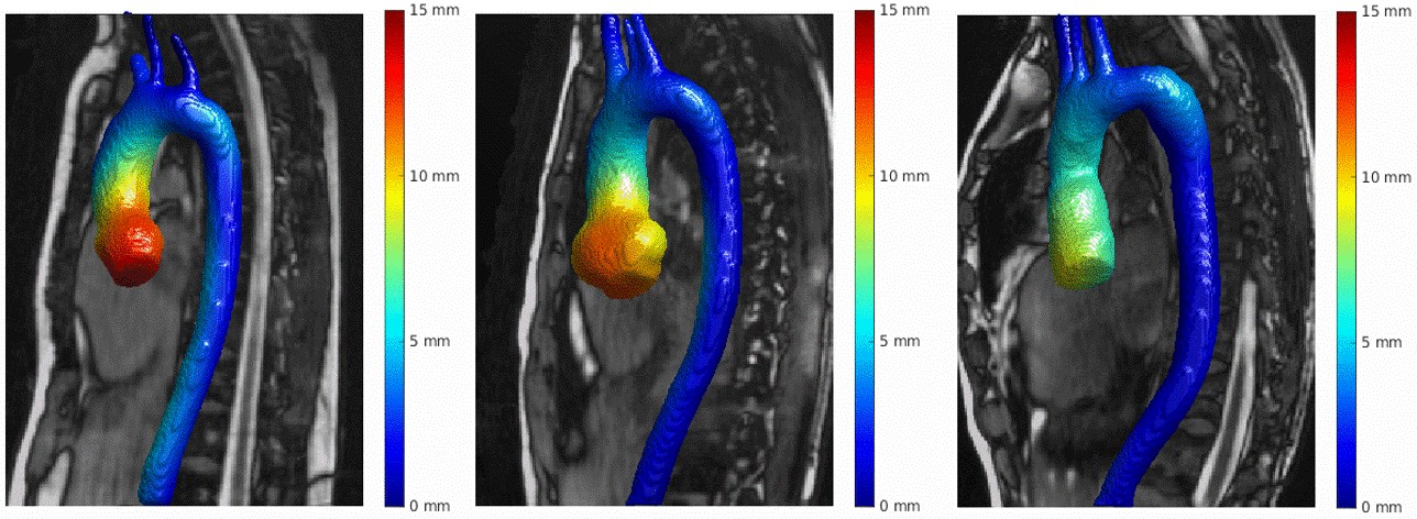 Figure 2: Quantification of aortic displacement over heartbeat. Left: healthy volunteer; center: Marfan patient who has not undergone surgery; right: Marfan patient after aortic root replacement surgery
Surgical Impact on Motion
Figure 2 shows that ascending aortic motion is reduced in surgically treated patients compared to healthy volunteers and non‑operated Marfan patients. This is expected: the implanted graft is stiffer than native aortic tissue and therefore restricts motion. Closer inspection reveals another pattern: proximal descending aortic motion is increased in operated patients relative to the other two groups. Co‑registration of aortas with abnormally increased motion demonstrated that 45% of operated patients (n=33) exhibit elevated motion in the proximal descending aorta (Figure 3).
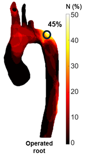 Figure 3. Quantification of aortic discplacement over the heartbeat
Energy Transfer Hypothesis
These unexpected findings raise an important biomechanical question: does the implanted graft redirect kinetic energy from blood flow in the ascending aorta toward the descending aorta? The team found a correlation between increased aortic motion and elevated blood flow velocities, measured using 4D flow MRI, in the proximal descending aortic segment. This suggests that altered energy distribution after graft implantation may drive localized increases in aortic wall motion, with potentially significant clinical implications.
Link to Type B Dissection
Marfan patients are known to have a higher risk of type B dissection following surgery. The newly observed increase in proximal descending aortic motion may provide a mechanistic explanation for this risk. In future research we can explore whether these biomechanical changes correlate directly with the occurrence of type B dissections, opening possibilities for improved risk stratification and personalized follow‑up strategies.
Evaluating the PEARS Procedure
At Amsterdam UMC, a different preventive operation is also frequently performed to avert type A dissection: the Personalized External Aortic Root Support (PEARS) procedure. In PEARS, a patient‑specific 3D‑printed external “mesh” is sewn around the aortic root. This support is more flexible than the conventional graft used in root replacement surgery. An important question is whether this increased flexibility reduces the transfer of blood flow energy toward the descending aorta, thereby limiting proximal descending motion and potentially lowering the risk of type B dissection.
Looking Ahead
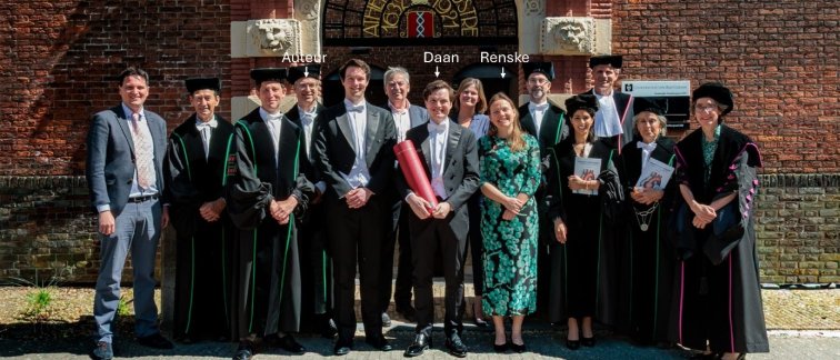
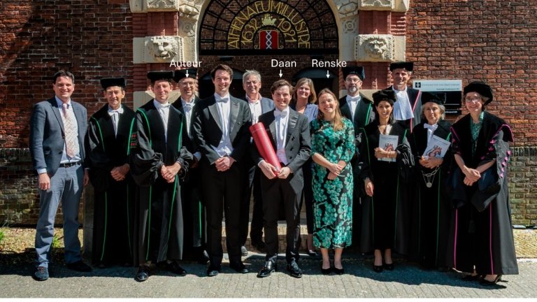
The technologies developed in this VIDI project, the high‑resolution 4D MRI of aortic motion, advanced segmentation, and motion quantification, offer powerful tools to investigate these questions. Although the formal project period has ended with Daan Bosshardt’s PhD defense in April 2026, the scientific collaboration continues. Renske now works as a postdoctoral researcher in Radiology & Nuclear Medicine, and Daan will start cardiology training in November 2026. Together, the team is well‑positioned to further explore how aortic biomechanics can guide safer, more personalized care for Marfan patients. For now, this marks a provisional end to a remarkable era in imaging aortic motion, but (depending on funds) also the beginning of new, clinically relevant research directions.
About the author: dr. ir. Pim van Ooij
Van Ooij is an Associate Professor and Principal Investigator in the Department of Radiology and Nuclear Medicine at Amsterdam UMC. He specializes in cardiovascular engineering and advanced MRI techniques. His research primarily focuses on the clinical translation of 4D (flow) MRI to map cardiovascular motion and blood flow patterns and improve the treatment of cardiovascular diseases.
He is affiliated with Amsterdam Cardiovascular Science (ACS), where he contributes as a member of Young ACS, a representative of the 2026 innovation Imaging and as a member of the Valorization Committee.