During Deep Brain Stimulation (DBS), two electrodes are implanted into deep brain structures. The tips of the electrodes deliver electrical pulses that help reduce symptoms such as tremors, stiffness, and slow movement.
Sharper images
However, not every patient responded equally well to DBS treatment. In some patients, motor symptoms improved less than expected. With the new 7-tesla MRI scanner, which uses a much stronger magnet than a conventional MRI and therefore produces higher-resolution images, each patient’s brain networks can be mapped in detail before surgery. This allows even deep brain structures and the connections between different brain regions to be visualized. “With this technique, we can determine much more precisely where to place the electrodes, specifically in the part of the brain structure involved in movement,” says neurosurgeon Maarten Bot of Amsterdam UMC. “As a result, we see a clear improvement in motor symptoms in a larger number of patients.”

Higher success rate
For the study, 102 Parkinson’s patients underwent surgery based on scans obtained with the new 7-tesla MRI scanner. Their outcomes were compared with those of 118 patients who had previously undergone DBS surgery using standard MRI images to determine electrode placement. Patients treated using the new method were more likely to achieve a clearly successful outcome. Among these patients, 96 percent experienced at least a 30 percent reduction in motor symptoms, compared with 86 percent in the standard-treatment group. The researchers assessed outcomes using a widely used Parkinson’s disease rating scale that measures the severity of motor symptoms. “For patients, the likelihood that the surgery will make a meaningful difference in daily life becomes greater. Symptoms such as tremors, stiffness, and slow movement improve more substantially,” says neuroscientist Yarit Wiggerts. “Ultimately, that is what we are striving for.”

A unique collaboration
Amsterdam UMC is the only hospital in the Netherlands authorized to use a 7-tesla MRI scanner, not only for research purposes but also clinically for patient care. This achievement follows years of collaboration among physicians, researchers, and technical specialists from Amsterdam UMC and the nearby Spinoza Centre for Neuroimaging, where the scanner is located. Their joint efforts led to approval of the scanner as an in-house developed medical device. According to the researchers, the study demonstrates the importance of collaboration between neuroscientists and clinicians. “This research truly emerged from the combination of technological innovation and direct patient care,” says Maarten Bot. “We can discuss clinical observations directly with neuroscientists and immediately apply those insights. That strengthens and accelerates the translation of new knowledge into the operating room.”
7-Tesla MRI for all patients
Most DBS procedures in the Netherlands are performed at Amsterdam UMC. From now on, all Parkinson’s disease patients undergoing this surgery will receive a 7-tesla MRI scan beforehand. For the time being, this technology is available only at Amsterdam UMC. However, the findings of this study may enable other DBS centers that have access to a 7-tesla MRI scanner to adopt the same approach quickly.
Read the publication in npj Parkinson’s Disease: Targeting based on patient-specific 7 Tesla MRI connectivity analysis improves deep brain stimulation for Parkinson’s disease
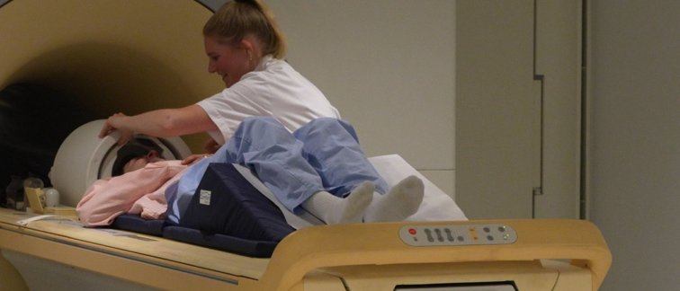
Image: Amsterdam UMC