

ACS proudly announces the PhD thesis of Fatimah Al Darwish, "Novel Imaging Approaches to Placental Insufficiency: Studies in the Reduced Uterine Perfusion Pressure rat model." Her work sheds light on how placental insufficiency develops over the course of pregnancy and shows how multimodal imaging can measure placental perfusion and oxygenation directly, identifying a critical window for early detection and intervention.
A clinical gap
Placental insufficiency, a failure to establish or maintain sufficient exchange between mother and fetus, underlies key obstetric complications such as preeclampsia and fetal growth restriction. Yet clinicians currently assess placental function indirectly, through fetal biometry and Doppler indices, and no widely effective therapy exists once insufficiency develops. "Clinically, we measure everything around the placenta but not the placenta itself. We look at fetal growth and blood flow patterns," Al Darwish explains. "But we can't directly see what's happening with oxygen or perfusion. That's what I wanted to address."

A multimodal toolbox
To study how placental insufficiency evolves, Al Darwish used the reduced uterine perfusion pressure (RUPP) rat model, in which uterine blood flow is mechanically reduced during pregnancy. She applied multiple imaging modalities: MRI (T2* mapping and 3D dynamic contrast-enhanced MRI), contrast-enhanced ultrasound, and photoacoustic imaging, complemented by histology of the maternal-fetal interface, the microscopic examination of tissue. "Using multiple imaging techniques together, we could measure both blood flow and oxygen directly in the living placenta. Then we looked at the actual tissue under the microscope to understand what the images were showing."
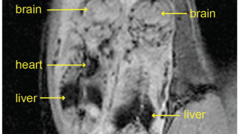
In vivo fetal MRI of a pregnant rat.
A story in time
Immediately after the reduction of uterine blood flow at gestational day 14, blood flow to the placenta was clearly impaired. At the same time, the uterine natural killer cells and trophoblasts that normally remodel the placenta's blood vessels did not migrate and invade as they should. By mid-gestation, perfusion and oxygenation appeared functionally compensated despite these structural abnormalities, and ultrasound and MRI readouts still correlated, even when differences between groups were small. By late gestation, blood flow measurements remained normal, while an MRI measure of placental oxygenation (T2*) indicated chronic hypoxia, suggesting that the lack of oxygen resulted from impaired remodeling of the placenta's blood vessels, rather than from insufficient blood flow alone. "The surprising part was seeing placentas that look functionally compensated but their structure wasn't remodeling normally," Al Darwish notes. " Then later, blood flow appears to recover, while the lack of oxygen only becomes visible near the end of pregnancy." Gestational days 14 to 15 emerge as a critical window for detecting acute perfusion impairment and evaluating early interventions. "That's the window where you can test if an early therapy works," she adds.
Hidden differences
Detailed analyses of the images, voxel by voxel, revealed marked differences between placentas within the same litter, with a subset deviating from normal pregnancy patterns. "Even within the same litter, placentas responded differently," Al Darwish explains. "Some clearly deviate from what you would see in a normal pregnancy, so you have to account for that, instead of only looking at group averages."
Towards time-specific monitoring
The blood flow problem, the abnormal development of the placenta's blood vessels, and the lack of oxygen do not all occur at the same time. Effective monitoring and treatment of placental insufficiency therefore require time-specific, multimodal imaging approaches that combine perfusion and oxygenation readouts and account for differences within and across placentas. Molecular tissue biomarkers are typically available only after delivery, and circulating biomarkers are proxies rather than direct readouts of placental function. That makes non-invasive imaging a promising approach to monitor placental function throughout pregnancy and to bridge between biological mechanisms and clinically relevant biomarkers. "You need the right imaging techniques at the right time.," Al Darwish concludes. "That is how you can test therapies in the window where they can still make a difference."
PhD defense
The public defense of Fatimah Al Darwish will take place on 8 October 2026, 10:00h at the Agnietenkapel. For more information, see the UvA website.