

Today, 29 September, is World Heart Day: a global initiative to raise awareness of cardiovascular disease. Amsterdam Cardiovascular Sciences marks this day by highlighting research that contributes to the prevention and treatment of cardiovascular disease. This year, the spotlight is on the ANOCA-CT study by Bettine van Willigen (Amsterdam UMC) and Fabienne Vervaat (Catharina Hospital Eindhoven), which aims to detect microvascular disease using solely the CT scan.
Chest pain, no answers
Insufficient blood supply to the heart muscle is a leading cause of morbidity and mortality worldwide. While obstructive coronary artery disease is the most common, a growing group of patients, mainly women, experience chest pain without detectable narrowing in the coronary arteries. This condition is known as Angina with No Obstructive Coronary Artery Disease (ANOCA). In some of these patients, the cause lies in the smallest blood vessels of the heart, the microvasculature, also referred to as microvascular disease.
An invasive diagnosis
Currently, this condition can be diagnosed using a Coronary Function Test (CFT), performed during cardiac catheterization. During a CFT, measurements are taken to assess both blood flow and resistance in the coronary arteries and the heart's microvasculature, including Coronary Flow Reserve (CFR) and the Index of Microvascular Resistance (IMR). However, CFT is an invasive, labor-intensive, and costly procedure with small but non-negligible risks, such as bleeding, stroke, allergic reactions, and coronary artery damage. In current practice, patients with chest pain first undergo a CT scan of the heart to non-invasively rule out obstructive coronary artery disease. If no abnormalities are found, medication is started pragmatically. Yet, many patients continue to experience long-term symptoms, and the next step is often a CFT. Due to the invasive nature of this diagnostic approach, the burden on the patient, and the cost, clinicians are reluctant to request a CFT. As a result, many patients remain without a diagnosis and live in uncertainty.
Team science
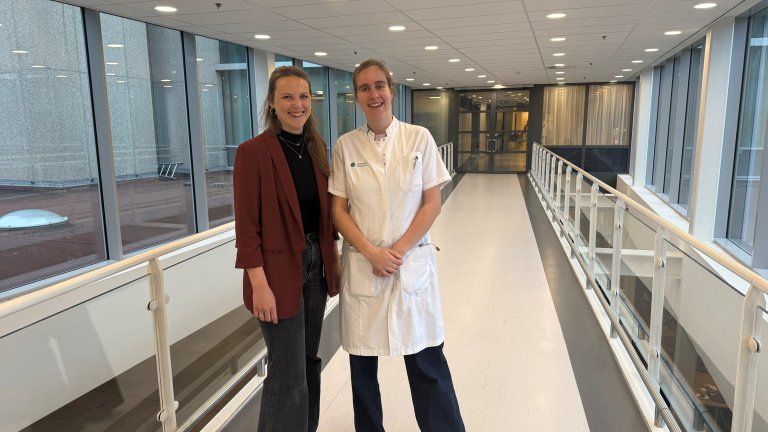
Cardiologist Fabienne Vervaat (Catharina Hospital Eindhoven) and Bettine van Willigen (postdoctoral researcher, Amsterdam UMC) received the Team Science Dekker Grant to develop a non-invasive detection method to detect microvascular disease using solely the CT scan. PhD candidate Ir. Hans Douben (TU/e) has since joined the team. In order to develop such a detection method, unraveling the underlying mechanisms of microvascular disease is a key element.
A synthetic microcirculation
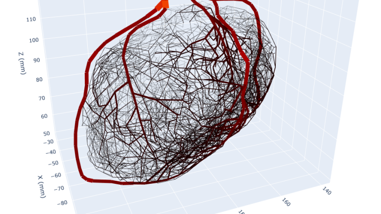
The team is developing a patient data-driven synthetic model of the left coronary microcirculation. The method consists of segmenting the coronary arteries from coronary CT angiography (CCTA) images. These vessels are then synthetically extended down to 10 μm arteriole radius size. "By introducing the structural and functional abnormalities that underlie microvascular disease, we can analyze in a controlled environment the effect of these abnormalities on the blood circulation in rest and hyperemia," explains Van Willigen. "This approach will provide insights into the mechanisms of microvascular disease, a first step toward a fully non-invasive detection method for microvascular disease."
The ANOCA-CT study is funded by the Dutch Heart Foundation through a Team Science Dekker Grant.