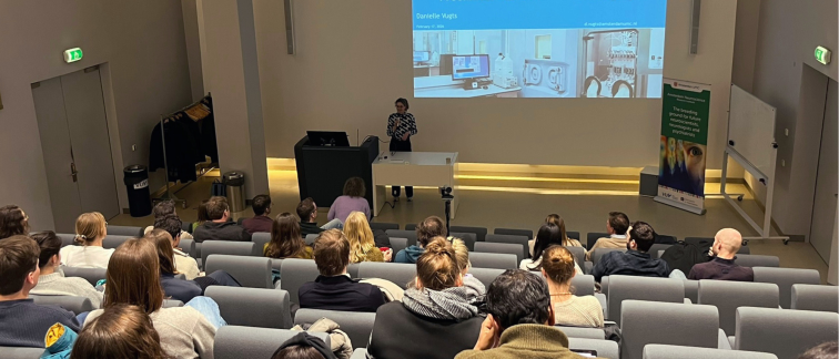
Danielle Vugts, Professor of PET Radiochemistry at Amsterdam UMC, presented on PET imaging using Zr-labelled antibodies. In recent years, this technique has become a valuable tool in drug development and may evolve into an essential de-risking strategy for immunotherapies targeting central nervous system diseases. A key question remains: can zirconium (Zr) labelling effectively image the localization and impact of biological drugs (such as immunotherapy) in the brain?
Vugts illustrated this challenge through the imaging of amyloid-beta plaques during aducanumab administration. Given the projected growth in the number of people living with dementia, studying drug distribution in the brain is of great importance. Vugts and her team aim to investigate whether further optimization of Zr chemistry is required and to develop a radiochemistry toolbox capable of labeling a wide range of biological drugs. This would create new opportunities to support drug development through advanced imaging.
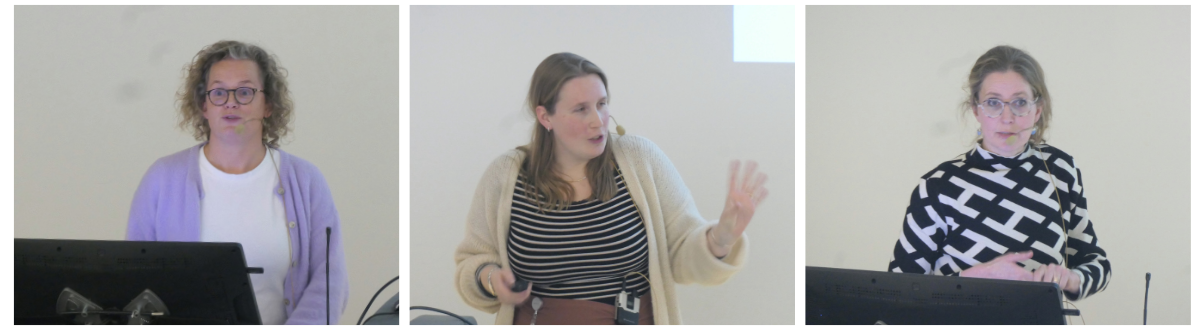
Lyduine Collij, Assistant Professor at Amsterdam UMC and the Alzheimer Center Amsterdam, discussed her views on the dynamic field of amyloid PET, and the transition from AMYPAD (Amyloid Imaging to Prevent Alzheimer’s Disease) to Euro-PAD, large consortia with multicenter data. AMYPAD represents the largest European longitudinal dataset phenotyping individuals at risk of Alzheimer’s disease progression. The consortium is now evolving into its next phase: the Euro-PAD collaborative framework.
By focusing on the preclinical and prodromal stages of Alzheimer’s disease, Euro-PAD aims to generate crucial insights into the earliest phases of the disease. Access to AMYPAD data is now available through the Alzheimer’s Disease Data Initiative. More information can be found at the AMYPAD website.
Collij also briefly elaborated on her awarded VENI project, titled “Advancing Amyloid-PET: Novel Biomarkers for Optimized Alzheimer’s Diagnosis and Risk Assessment.” She emphasized that amyloid-PET scans contain a wealth of information and highlighted the importance of collaboration and data sharing to fully unlock their potential.
Finally, Elsmarieke van de Giessen, radiologist at Amsterdam UMC, built on the previous presentations by emphasizing the added value of combining imaging measures with other modalities, such as proteomics. She presented work on tau-PET imaging combined with proteomic analyses, demonstrating how multimodal approaches can provide deeper biological insight.
Van de Giessen also elaborated on her VIDI project, “The neuroinflammatory process in Alzheimer’s disease.” Her research focuses on characterizing the neuroinflammatory process and developing measurable markers to determine its different phases. Ultimately, she aims to create a roadmap linking specific brain regions to neuroinflammation-related proteins. This approach could help identify the right patients for the right treatment in the future.
A special thanks goes to Elsmarieke van de Giessen for hosting this successful event. The next Brain Imaging Symposium will be organized at the beginning of the summer.
The Brain Imaging research program is one of the nine research programs within Amsterdam Neuroscience, led by Menno Schoonheim, Matthan Caan, Elsmarieke van de Giessen, and Martijn Beudel. This program aims to make a difference in the field of neuroimaging, as well as provide services to other medical disciplines, through the optimization of infrastructure, analyses, techniques and technologies.